Heel Aches The Primary Causes, Signals And Treatment Methods
Overview

Heel pain is most often caused by plantar fasciitis, a condition that is sometimes also called heel spur syndrome when a spur is present. Heel pain may also be due to other causes, such as a stress fracture, tendonitis, arthritis, nerve irritation, or rarely, a cyst. Because there are several potential causes, it is important to have heel pain properly diagnosed. A foot and ankle surgeon is able to distinguish between all the possibilities and determine the underlying source of your heel pain.
Causes
The most common cause of heel pain is plantar fasciitis. Plantar fasciitis is a stretching of the plantar fascia, a ligament that runs from the ball of foot through the arch and is attached to the heel. It is that attachment which becomes aggravated and typically causes pain after being on your feet for lengths of time. Abnormal motion of the foot (pronation) is one cause of plantar fasciitis. Heel spurs, which are abnormal bone growths coming off the heel, can also cause heel pain. Other causes include repetitive stress or shock to the heel, standing for prolonged periods or osteoarthritis.
Symptoms
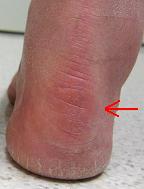
Sever?s Disease. This is a condition that occurs in 10 - 15 year old children, predominantly boys and is associated with running and repetitive jumping. It is also associated with flimsy footwear that kids may wear. It occurs when the Achilles tendon continually pulls on the apophysis of the calcaneum and does not allow for it to fuse with the body of the calcaneum. Calcaneal enthesopathy. This occurs when there is repetitive trauma at the attachment of the Achilles tendon, resulting in a spur from the calcaneum up into the Achilles tendon. It is usually visualized on x-ray and may be tender if there is an associated bursitis or tendonitis. "Pump Bump". Also known as Haglund?s Deformity, this is a bony enlargement that exists on the back of the heel - usually related to a congenital abnormality or with chronic bursitis, causing a thickening. There may have already been trauma or pressure from footwear. Treatment is usually protection of the bump and correct footwear. Associated with a symmetrical swelling at the base of the Achilles tendon. It is usually related to repetitive trauma or inappropriate footwear. It is often red and hot in the early stages. Treatment is usually to correct the footwear, provide padding and treat the local symptoms e.g. ice, rest, physiotherapy and cortisone injection. Fat Pad Syndrome. Direct contact with the base of the heel may result in trauma to the fat pad. Related to obesity, training on hard surfaces, uneven grounds, poor shoes especially overlarge shoes which can cause shearing forces on the heel. These conditions are renowned for taking a long time to recover - usually many months.
Diagnosis
Your GP or podiatrist (a healthcare professional who specialises in foot care) may be able to diagnose the cause of your heel pain by asking about your symptoms and examining your heel and foot. You will usually only need further tests if you have additional symptoms that suggest the cause of your heel pain is not inflammation, such as numbness or a tingling sensation in your foot - this could be a sign of nerve damage in your feet and legs (peripheral neuropathy), your foot feels hot and you have a high temperature (fever) of 38C (100.4F) or above - these could be signs of a bone infection, you have stiffness and swelling in your heel - this could be a sign of arthritis. Possible further tests may include, blood tests, X-rays - where small doses of radiation are used to detect problems with your bones and tissues, a magnetic resonance imaging (MRI) scan or ultrasound scan, which are more detailed scans.
Non Surgical Treatment
Treatment of plantar fasciitis is usually performed in stages according to the duration and degree of pain. Treatment may take many months if the condition has been longstanding. Treatment usually begins with anti-inflammatory medication, shoe modification, temporary limitation of activities, weight loss and heel cord stretching. Also, night splints are often helpful to stretch the plantar fascia. An arch support (orthotic) may also be helpful, especially if you have a flat foot. If the problem continues, the tender area occasionally may be injected with cortisone and a local anesthetic. For a difficult, chronic problem, a period of casting may be used to improve this condition. Surgical treatment is rarely needed. If performed, it aims to partially release the plantar fascia and stimulate healing of the chronic inflammation. Removal of a heel spur, if it is large, may also be done at the time of surgery.
Surgical Treatment
When a diagnosis of plantar fasciitis is made early, most patients respond to conservative treatment and don?t require surgical intervention. Often, when there is a secondary diagnosis contributing to your pain, such as an entrapped nerve, and you are non-responsive to conservative care, surgery may be considered. Dr. Talarico will discuss all options and which approach would be the most beneficial for your condition.
foot pain heel
Prevention
Wear shoes that fit well, front, back and sides and have shock-absorbent soles, rigid uppers and supportive heel counters. Do not wear shoes with excessive wear on heels or soles. Prepare properly before exercising. Warm-up before running or walking, and do some stretching exercises afterward. Pace yourself when you participate in athletic activities. If overweight, try non weight-bearing activities such as swimming or cycling. Your podiatrist may also use taping or strapping to provide extra support for your foot. Orthoses (shoe inserts) specifically made to suit your needs may be also be prescribed.
Heel pain is most often caused by plantar fasciitis, a condition that is sometimes also called heel spur syndrome when a spur is present. Heel pain may also be due to other causes, such as a stress fracture, tendonitis, arthritis, nerve irritation, or rarely, a cyst. Because there are several potential causes, it is important to have heel pain properly diagnosed. A foot and ankle surgeon is able to distinguish between all the possibilities and determine the underlying source of your heel pain.
Causes
The most common cause of heel pain is plantar fasciitis. Plantar fasciitis is a stretching of the plantar fascia, a ligament that runs from the ball of foot through the arch and is attached to the heel. It is that attachment which becomes aggravated and typically causes pain after being on your feet for lengths of time. Abnormal motion of the foot (pronation) is one cause of plantar fasciitis. Heel spurs, which are abnormal bone growths coming off the heel, can also cause heel pain. Other causes include repetitive stress or shock to the heel, standing for prolonged periods or osteoarthritis.
Symptoms
Sever?s Disease. This is a condition that occurs in 10 - 15 year old children, predominantly boys and is associated with running and repetitive jumping. It is also associated with flimsy footwear that kids may wear. It occurs when the Achilles tendon continually pulls on the apophysis of the calcaneum and does not allow for it to fuse with the body of the calcaneum. Calcaneal enthesopathy. This occurs when there is repetitive trauma at the attachment of the Achilles tendon, resulting in a spur from the calcaneum up into the Achilles tendon. It is usually visualized on x-ray and may be tender if there is an associated bursitis or tendonitis. "Pump Bump". Also known as Haglund?s Deformity, this is a bony enlargement that exists on the back of the heel - usually related to a congenital abnormality or with chronic bursitis, causing a thickening. There may have already been trauma or pressure from footwear. Treatment is usually protection of the bump and correct footwear. Associated with a symmetrical swelling at the base of the Achilles tendon. It is usually related to repetitive trauma or inappropriate footwear. It is often red and hot in the early stages. Treatment is usually to correct the footwear, provide padding and treat the local symptoms e.g. ice, rest, physiotherapy and cortisone injection. Fat Pad Syndrome. Direct contact with the base of the heel may result in trauma to the fat pad. Related to obesity, training on hard surfaces, uneven grounds, poor shoes especially overlarge shoes which can cause shearing forces on the heel. These conditions are renowned for taking a long time to recover - usually many months.
Diagnosis
Your GP or podiatrist (a healthcare professional who specialises in foot care) may be able to diagnose the cause of your heel pain by asking about your symptoms and examining your heel and foot. You will usually only need further tests if you have additional symptoms that suggest the cause of your heel pain is not inflammation, such as numbness or a tingling sensation in your foot - this could be a sign of nerve damage in your feet and legs (peripheral neuropathy), your foot feels hot and you have a high temperature (fever) of 38C (100.4F) or above - these could be signs of a bone infection, you have stiffness and swelling in your heel - this could be a sign of arthritis. Possible further tests may include, blood tests, X-rays - where small doses of radiation are used to detect problems with your bones and tissues, a magnetic resonance imaging (MRI) scan or ultrasound scan, which are more detailed scans.
Non Surgical Treatment
Treatment of plantar fasciitis is usually performed in stages according to the duration and degree of pain. Treatment may take many months if the condition has been longstanding. Treatment usually begins with anti-inflammatory medication, shoe modification, temporary limitation of activities, weight loss and heel cord stretching. Also, night splints are often helpful to stretch the plantar fascia. An arch support (orthotic) may also be helpful, especially if you have a flat foot. If the problem continues, the tender area occasionally may be injected with cortisone and a local anesthetic. For a difficult, chronic problem, a period of casting may be used to improve this condition. Surgical treatment is rarely needed. If performed, it aims to partially release the plantar fascia and stimulate healing of the chronic inflammation. Removal of a heel spur, if it is large, may also be done at the time of surgery.
Surgical Treatment
When a diagnosis of plantar fasciitis is made early, most patients respond to conservative treatment and don?t require surgical intervention. Often, when there is a secondary diagnosis contributing to your pain, such as an entrapped nerve, and you are non-responsive to conservative care, surgery may be considered. Dr. Talarico will discuss all options and which approach would be the most beneficial for your condition.
foot pain heel
Prevention
Wear shoes that fit well, front, back and sides and have shock-absorbent soles, rigid uppers and supportive heel counters. Do not wear shoes with excessive wear on heels or soles. Prepare properly before exercising. Warm-up before running or walking, and do some stretching exercises afterward. Pace yourself when you participate in athletic activities. If overweight, try non weight-bearing activities such as swimming or cycling. Your podiatrist may also use taping or strapping to provide extra support for your foot. Orthoses (shoe inserts) specifically made to suit your needs may be also be prescribed.
The Way To Fix Functional Leg Length Discrepancy
Overview
Surgeries for LLD are designed to do one of three general things ? shorten the long leg, stop or slow the growth of the longer or more rapidly growing leg, or lengthen the short leg. Stopping the growth of the longer leg is the most commonly utilized of the three approaches and involves an operation known as an epiphysiodesis , in which the growth plate of either the lower femur or upper tibia is visualized in the operating room using fluoroscopy (a type of real-time radiographic imaging) and ablated , which involves drilling into the region several times, such that the tissue is no longer capable of bone growth. Because the epiphyseal growth capabilities cannot be restored following the surgery, proper timing is crucial. Usually the operation is planned for the last 2 to 3 years of growth and has excellent results, with children leaving the hospital within a few days with good mobility. However, it is only appropriate for LLD of under 5cm.
Causes
There are many causes of leg length discrepancy. Some include, A broken leg bone may lead to a leg length discrepancy if it heals in a shortened position. This is more likely if the bone was broken in many pieces. It also is more likely if skin and muscle tissue around the bone were severely injured and exposed, as in an open fracture. Broken bones in children sometimes grow faster for several years after healing, causing the injured bone to become longer. A break in a child's bone through the growth center near the end of the bone may cause slower growth, resulting in a shorter leg. Bone infections that occur in children while they are growing may cause a significant leg length discrepancy. This is especially true if the infection happens in infancy. Inflammation of joints during growth may cause unequal leg length. One example is juvenile arthritis. Bone diseases may cause leg length discrepancy, as well. Examples are, Neurofibromatosis, Multiple hereditary exostoses, Ollier disease. Other causes include inflammation (arthritis) and neurologic conditions. Sometimes the cause of leg length discrepancy is unknown, particularly in cases involving underdevelopment of the inner or outer side of the leg, or partial overgrowth of one side of the body. These conditions are usually present at birth, but the leg length difference may be too small to be detected. As the child grows, the leg length discrepancy increases and becomes more noticeable. In underdevelopment, one of the two bones between the knee and the ankle is abnormally short. There also may be related foot or knee problems. Hemihypertrophy (one side too big) or hemiatrophy (one side too small) are rare leg length discrepancy conditions. In these conditions, the arm and leg on one side of the body are either longer or shorter than the arm and leg on the other side of the body. There may also be a difference between the two sides of the face. Sometimes no cause can be found. This is known as an "idiopathic" difference.
Symptoms
The symptoms of limb deformity can range from a mild difference in the appearance of a leg or arm to major loss of function of the use of an extremity. For instance, you may notice that your child has a significant limp. If there is deformity in the extremity, the patient may develop arthritis as he or she gets older, especially if the lower extremities are involved. Patients often present due to the appearance of the extremity (it looks different from the other side).
Diagnosis
Infants, children or adolescents suspected of having a limb-length condition should receive an evaluation at the first sign of difficulty in using their arms or legs. In many cases, signs are subtle and only noticeable in certain situations, such as when buying clothing or playing sports. Proper initial assessments by qualified pediatric orthopedic providers can reduce the likelihood of long-term complications and increase the likelihood that less invasive management will be effective. In most cases, very mild limb length discrepancies require no formal treatment at all.
Non Surgical Treatment
For minor limb length discrepancy in patients with no deformity, treatment may not be necessary. Because the risks may outweigh the benefits, surgical treatment to equalize leg lengths is usually not recommended if the difference is less than 1 inch. For these small differences, the physician may recommend a shoe lift. A lift fitted to the shoe can often improve walking and running, as well as relieve any back pain that may be caused by the limb length discrepancy. Shoe lifts are inexpensive and can be removed if they are not effective.

height increase food
Surgical Treatment
Limb deformity or leg length problems can be treated by applying an external frame to the leg. The frame consists of metal rings which go round the limb. The rings are held onto the body by wires and metal pins which pass through the skin and are anchored into the bone. During this operation, the bone is divided. Gradual adjustment of the frame results in creation of a new bone allowing a limb to be lengthened. The procedure involves the child having an anaesthetic. The child is normally in hospital for one week. The child and family are encouraged to clean pin sites around the limb. The adjustments of the frame (distractions) are performed by the child and/or family. The child is normally encouraged to walk on the operated limb and to actively exercise the joints above and below the frame. The child is normally reviewed on a weekly basis in clinic to monitor the correction of the deformity. The frame normally remains in place for 3 months up to one year depending on the condition which is being treated. The frame is normally removed under a general anaesthetic at the end of treatment.
Surgeries for LLD are designed to do one of three general things ? shorten the long leg, stop or slow the growth of the longer or more rapidly growing leg, or lengthen the short leg. Stopping the growth of the longer leg is the most commonly utilized of the three approaches and involves an operation known as an epiphysiodesis , in which the growth plate of either the lower femur or upper tibia is visualized in the operating room using fluoroscopy (a type of real-time radiographic imaging) and ablated , which involves drilling into the region several times, such that the tissue is no longer capable of bone growth. Because the epiphyseal growth capabilities cannot be restored following the surgery, proper timing is crucial. Usually the operation is planned for the last 2 to 3 years of growth and has excellent results, with children leaving the hospital within a few days with good mobility. However, it is only appropriate for LLD of under 5cm.
Causes
There are many causes of leg length discrepancy. Some include, A broken leg bone may lead to a leg length discrepancy if it heals in a shortened position. This is more likely if the bone was broken in many pieces. It also is more likely if skin and muscle tissue around the bone were severely injured and exposed, as in an open fracture. Broken bones in children sometimes grow faster for several years after healing, causing the injured bone to become longer. A break in a child's bone through the growth center near the end of the bone may cause slower growth, resulting in a shorter leg. Bone infections that occur in children while they are growing may cause a significant leg length discrepancy. This is especially true if the infection happens in infancy. Inflammation of joints during growth may cause unequal leg length. One example is juvenile arthritis. Bone diseases may cause leg length discrepancy, as well. Examples are, Neurofibromatosis, Multiple hereditary exostoses, Ollier disease. Other causes include inflammation (arthritis) and neurologic conditions. Sometimes the cause of leg length discrepancy is unknown, particularly in cases involving underdevelopment of the inner or outer side of the leg, or partial overgrowth of one side of the body. These conditions are usually present at birth, but the leg length difference may be too small to be detected. As the child grows, the leg length discrepancy increases and becomes more noticeable. In underdevelopment, one of the two bones between the knee and the ankle is abnormally short. There also may be related foot or knee problems. Hemihypertrophy (one side too big) or hemiatrophy (one side too small) are rare leg length discrepancy conditions. In these conditions, the arm and leg on one side of the body are either longer or shorter than the arm and leg on the other side of the body. There may also be a difference between the two sides of the face. Sometimes no cause can be found. This is known as an "idiopathic" difference.
Symptoms
The symptoms of limb deformity can range from a mild difference in the appearance of a leg or arm to major loss of function of the use of an extremity. For instance, you may notice that your child has a significant limp. If there is deformity in the extremity, the patient may develop arthritis as he or she gets older, especially if the lower extremities are involved. Patients often present due to the appearance of the extremity (it looks different from the other side).
Diagnosis
Infants, children or adolescents suspected of having a limb-length condition should receive an evaluation at the first sign of difficulty in using their arms or legs. In many cases, signs are subtle and only noticeable in certain situations, such as when buying clothing or playing sports. Proper initial assessments by qualified pediatric orthopedic providers can reduce the likelihood of long-term complications and increase the likelihood that less invasive management will be effective. In most cases, very mild limb length discrepancies require no formal treatment at all.
Non Surgical Treatment
For minor limb length discrepancy in patients with no deformity, treatment may not be necessary. Because the risks may outweigh the benefits, surgical treatment to equalize leg lengths is usually not recommended if the difference is less than 1 inch. For these small differences, the physician may recommend a shoe lift. A lift fitted to the shoe can often improve walking and running, as well as relieve any back pain that may be caused by the limb length discrepancy. Shoe lifts are inexpensive and can be removed if they are not effective.
height increase food
Surgical Treatment
Limb deformity or leg length problems can be treated by applying an external frame to the leg. The frame consists of metal rings which go round the limb. The rings are held onto the body by wires and metal pins which pass through the skin and are anchored into the bone. During this operation, the bone is divided. Gradual adjustment of the frame results in creation of a new bone allowing a limb to be lengthened. The procedure involves the child having an anaesthetic. The child is normally in hospital for one week. The child and family are encouraged to clean pin sites around the limb. The adjustments of the frame (distractions) are performed by the child and/or family. The child is normally encouraged to walk on the operated limb and to actively exercise the joints above and below the frame. The child is normally reviewed on a weekly basis in clinic to monitor the correction of the deformity. The frame normally remains in place for 3 months up to one year depending on the condition which is being treated. The frame is normally removed under a general anaesthetic at the end of treatment.
Working with Mortons Neuroma
Overview
 A Morton?s Neuroma is actually incorrectly termed, with the name suggesting it is a tumour or growth. Rather than a true neuroma it is actually what is called a perineural fibrosis, which means that over time the sheath surrounding the nerve becomes irritated, inflamed, and forms a thickened scar tissue.
Causes
There are a number of common causes for Morton?s Neuroma, (though the condition can arise spontaneously for reasons still unknown). The Neuroma often occurs in response to irritation, pressure or traumatic injury to one of the digital nerves leading to the toes. A thickening of nerve tissue results as part of the body?s response to the irritation or injury. Abnormal foot movement used to compensate for bunions, hammertoes, flatfeet and other conditions can lead to irritation and development of Morton?s Neuroma. Pronation of the foot may cause the heads of the metatarsal bones to rotate slightly, thereby pinching the nerve running between the metatarsal heads. Chronic pressure or pinching causes the nerve sheath to enlarge, becoming increasingly squeezed, producing worsening pain over time, if not addressed. Morton?s Neuroma can be exacerbated when tight shoes providing little room for the forefoot are worn. Activities which over-pronate the foot (such as walking barefoot in sand) may increase the pain associated with Morton?s Neuroma, as will any high-impact activity, such as jogging.
Symptoms
People with Morton's neuroma usually complain of pain that can start in the ball of the foot and shoot into the affected toes. However, some people just have toe pain. There may also be burning and tingling of the toes. The symptoms are usually felt up the sides of the space between two toes. For example, if the nerve between the third and fourth long bones (metatarsals) of the right foot is affected, the symptoms will usually be felt up the right-hand side of the fourth toe and up the left-hand side of the third toe. Some people describe the pain that they feel as being like walking on a stone or a marble. Symptoms can be made worse if you wear high-heeled shoes. The pain is relieved by taking your shoe off, resting your foot and massaging the area. You may also experience some numbness between the affected toes. Your affected toes may also appear to be spread apart, which doctors refer to as the 'V sign'. The symptoms can vary and may come and go over a number of years. For example, some people may experience two attacks of pain in a week and then nothing for a year. Others may have regular and persistent (chronic) pain.
Diagnosis
In some cases your doctor will be able to feel the Morton's as a swelling in the middle of your foot. However they may also suggest an X-ray or a blood test - this is normally to rule our other causes of the pain such as arthritis. The most accurate way to diagnose Morton?s itself is with magnetic resonance imaging (MRI) or ultrasound.
Non Surgical Treatment
Ice therapy and anti-inflammatory medications or supplements. If conservative care measures fail to resolve your problem, some foot care providers may recommend a cortisone injection around your involved nerve to help reduce your swelling and inflammation. Concentrated alcohol injections around your affected nerve have also shown good results and should be considered before undergoing neurectomy, a surgical procedure to remove the enlarged, traumatized portion of your involved nerve.
Surgical Treatment
Surgery. This is the last and most permanent course of action. This surgery is used as a last resort as it often comes with a series of side affects including the risk of making the pain worse. This surgery can be performed by Orthopedic surgeons as well as Podiatric surgeons.
A Morton?s Neuroma is actually incorrectly termed, with the name suggesting it is a tumour or growth. Rather than a true neuroma it is actually what is called a perineural fibrosis, which means that over time the sheath surrounding the nerve becomes irritated, inflamed, and forms a thickened scar tissue.Causes
There are a number of common causes for Morton?s Neuroma, (though the condition can arise spontaneously for reasons still unknown). The Neuroma often occurs in response to irritation, pressure or traumatic injury to one of the digital nerves leading to the toes. A thickening of nerve tissue results as part of the body?s response to the irritation or injury. Abnormal foot movement used to compensate for bunions, hammertoes, flatfeet and other conditions can lead to irritation and development of Morton?s Neuroma. Pronation of the foot may cause the heads of the metatarsal bones to rotate slightly, thereby pinching the nerve running between the metatarsal heads. Chronic pressure or pinching causes the nerve sheath to enlarge, becoming increasingly squeezed, producing worsening pain over time, if not addressed. Morton?s Neuroma can be exacerbated when tight shoes providing little room for the forefoot are worn. Activities which over-pronate the foot (such as walking barefoot in sand) may increase the pain associated with Morton?s Neuroma, as will any high-impact activity, such as jogging.
Symptoms
People with Morton's neuroma usually complain of pain that can start in the ball of the foot and shoot into the affected toes. However, some people just have toe pain. There may also be burning and tingling of the toes. The symptoms are usually felt up the sides of the space between two toes. For example, if the nerve between the third and fourth long bones (metatarsals) of the right foot is affected, the symptoms will usually be felt up the right-hand side of the fourth toe and up the left-hand side of the third toe. Some people describe the pain that they feel as being like walking on a stone or a marble. Symptoms can be made worse if you wear high-heeled shoes. The pain is relieved by taking your shoe off, resting your foot and massaging the area. You may also experience some numbness between the affected toes. Your affected toes may also appear to be spread apart, which doctors refer to as the 'V sign'. The symptoms can vary and may come and go over a number of years. For example, some people may experience two attacks of pain in a week and then nothing for a year. Others may have regular and persistent (chronic) pain.
Diagnosis
In some cases your doctor will be able to feel the Morton's as a swelling in the middle of your foot. However they may also suggest an X-ray or a blood test - this is normally to rule our other causes of the pain such as arthritis. The most accurate way to diagnose Morton?s itself is with magnetic resonance imaging (MRI) or ultrasound.
Non Surgical Treatment
Ice therapy and anti-inflammatory medications or supplements. If conservative care measures fail to resolve your problem, some foot care providers may recommend a cortisone injection around your involved nerve to help reduce your swelling and inflammation. Concentrated alcohol injections around your affected nerve have also shown good results and should be considered before undergoing neurectomy, a surgical procedure to remove the enlarged, traumatized portion of your involved nerve.
Surgical Treatment
Surgery. This is the last and most permanent course of action. This surgery is used as a last resort as it often comes with a series of side affects including the risk of making the pain worse. This surgery can be performed by Orthopedic surgeons as well as Podiatric surgeons.
Shoe Lifts The Experts Option For Leg Length Discrepancy
There are actually two unique variations of leg length discrepancies, congenital and acquired. Congenital indicates you are born with it. One leg is structurally shorter compared to the other. As a result of developmental phases of aging, the brain picks up on the stride pattern and recognizes some variation. Our bodies usually adapts by tilting one shoulder to the "short" side. A difference of less than a quarter inch isn't grossly uncommon, demand Shoe Lifts to compensate and normally does not have a profound effect over a lifetime.

Leg length inequality goes mainly undiagnosed on a daily basis, yet this condition is easily solved, and can eradicate numerous instances of lower back pain.
Therapy for leg length inequality commonly consists of Shoe Lifts. These are low-priced, usually priced at below twenty dollars, compared to a custom orthotic of $200 or higher. When the amount of leg length inequality begins to exceed half an inch, a whole sole lift is generally the better choice than a heel lift. This prevents the foot from being unnecessarily stressed in an abnormal position.
Back pain is easily the most prevalent condition impacting people today. Over 80 million people have problems with back pain at some stage in their life. It's a problem which costs employers vast amounts of money year after year due to time lost and productivity. Fresh and better treatment solutions are constantly sought after in the hope of minimizing the economic impact this issue causes.

Men and women from all corners of the earth experience foot ache due to leg length discrepancy. In these types of situations Shoe Lifts are usually of very useful. The lifts are capable of alleviating any discomfort and pain in the feet. Shoe Lifts are recommended by countless qualified orthopaedic physicians.
So as to support the human body in a healthy and balanced fashion, feet have got a critical job to play. Irrespective of that, it is sometimes the most overlooked area in the body. Many people have flat-feet which means there may be unequal force placed on the feet. This will cause other body parts like knees, ankles and backs to be affected too. Shoe Lifts make sure that proper posture and balance are restored.
Leg length inequality goes mainly undiagnosed on a daily basis, yet this condition is easily solved, and can eradicate numerous instances of lower back pain.
Therapy for leg length inequality commonly consists of Shoe Lifts. These are low-priced, usually priced at below twenty dollars, compared to a custom orthotic of $200 or higher. When the amount of leg length inequality begins to exceed half an inch, a whole sole lift is generally the better choice than a heel lift. This prevents the foot from being unnecessarily stressed in an abnormal position.
Back pain is easily the most prevalent condition impacting people today. Over 80 million people have problems with back pain at some stage in their life. It's a problem which costs employers vast amounts of money year after year due to time lost and productivity. Fresh and better treatment solutions are constantly sought after in the hope of minimizing the economic impact this issue causes.
Men and women from all corners of the earth experience foot ache due to leg length discrepancy. In these types of situations Shoe Lifts are usually of very useful. The lifts are capable of alleviating any discomfort and pain in the feet. Shoe Lifts are recommended by countless qualified orthopaedic physicians.
So as to support the human body in a healthy and balanced fashion, feet have got a critical job to play. Irrespective of that, it is sometimes the most overlooked area in the body. Many people have flat-feet which means there may be unequal force placed on the feet. This will cause other body parts like knees, ankles and backs to be affected too. Shoe Lifts make sure that proper posture and balance are restored.
What May Cause Heel Spur

Overview
Heel spurs are a relatively common cause of heel pain. A heel spur is a pointed bone fragment that extends forward from the bottom of the heel from the heel bone, also referred to as a calcaneous. Serious pain and discomfort often develops with this condition. In many cases, a heel spur develops along with plantar fasciitis which occurs when the plantar fascia ligament becomes inflamed.
Causes
Faulty foot structures such as abnormal growths, different leg lengths, and unhealed injuries and haveinf flat feet or high arches. Muscle imbalances tight, weak or shortened muscles in your foot, plantar fascia, ankle, calf and hamstring. Over pronation can cause imbalance in foot mechanics which puts excess pressure on the plantar fascia. Poor biomechanics affect the way your foot hits the ground. If you overpronate (feet roll inward) you tend to have flat feet (pes planus), which increases stress on the heel bone. Regular shoes or high heels that are too tight or don't support your heel or arch affect the distribution of your body weight on your foot. Health conditions such as obesity, inflammatory diseases (rheumatoid arthritis, ankylosing spondylitis), bursitis, neuroma (nerve growths), gout, diabetes, Haglund's deformity, and Achilles tendinitis can also instigate the problem. Running or jogging on hard surfaces, repetative striking of the heel bone.

Symptoms
Heel spurs can be quite painful, but can just as likely occur with no symptoms at all. Plantar fasciitis is a contributing condition to heel spurs. The cause of the pain is not the heel spur itself but the soft-tissue injury associated with it. The feeling has been described as a knife or pin sticking into the bottom of your feet when you first stand up after sitting or laying down for a long period of time - a pain that later turns into a dull ache.
Diagnosis
A heel spur is often seen on X-ray as a bony protrusion, which can vary in size. However, because a Heel Spur only indicates increased load on the plantar fascia, and not pain, an ultra sound may be required to assess other actual cause of the heel pain such and may include checking to see if the plantar fascia is inflamed or degenerated.
Non Surgical Treatment
In some cases, heel spur pain may not be resolved through conservative treatment options. In those cases, cortisone injections may be used to reduce inflammation associated with the condition, helping to reduce discomfort. However, treatment options such as these must be discussed in detail with your physician, since more serious forms of treatment could yield negative side effects, such as atrophy of the heel's fat pad, or the rupture of the plantar fascia ligament. Although such side effects are rare, they are potential problems that could deliver added heel pain.
Surgical Treatment
Heel spur surgery should only be considered after less invasive treatment methods have been explored and ruled insufficient. The traditional surgical approach to treating heel spurs requires a scalpel cut to the bottom of the food which allows the surgeon to access the bone spur. Endoscopic plantar fasciotomies (EPF) involve one or two small incisions in the foot which allow the surgeon to access and operate on the bone spur endoscopically. Taking a surgical approach to heel spur treatment is a topic to explore with a foot and ankle specialist.
Posterior Calcaneal Spur Causes

Overview
Heel spurs are usually under the heel and are generally caused by excessive forces acting on the bone. By far the most common cause of heel spurs is abnormal biomechanics - often the same biomechanics that cause plantar fasciitis. Heel spurs are not a direct cause of heel pain. They grow in response to the forces of the soft tissue pulling on the bone. Any condition where the foot has excessive motion can produce tension within the soft tissues acting on the heel.
Causes
Causes for heel spurs (and related plantar fasciitis) include increase or change in activity, lack of arch support or poor shoe choice, injury, inflexibility in Achilles tendon and calf muscles, and spending hours daily on the feet. Also, arthritis from aging is often a common cause of bone loss and natural cushioning under the heel. Tarsal tunnel syndrome can also be to blame. Ultimately, in the United States, the most likely cause of this pain is being overweight. With more than 60% of the nation obese or morbidly obese, foot pain related to excessive weight is most likely. Dietary changes are most likely to cause long-term relief for bone spurs and plantar fasciitis.

Symptoms
Pain and discomfort associated with heel spurs does not occur from the spur itself. The bone growth itself has no feeling. However, as you move, this growth digs into sensitive nerves and tissue along the heel of the foot, resulting in severe pain. Pain can also be generated when pushing off with the toes while walking. Swelling along the heel is also common.
Diagnosis
Your doctor, when diagnosing and treating this condition will need an x-ray and sometimes a gait analysis to ascertain the exact cause of this condition. If you have pain in the bottom of your foot and you do not have diabetes or a vascular problem, some of the over-the-counter anti-inflammatory products such as Advil or Ibuprofin are helpful in eradicating the pain. Pain creams, such as Neuro-eze, BioFreeze & Boswella Cream can help to relieve pain and help increase circulation.
Non Surgical Treatment
Bone spurs rarely require treatment unless they are causing frequent pain or damaging other tissues. Because heel spurs and plantar fasciitis are so closely related, they are usually treated the same way. Symptomatic treatment involves rest, especially from the activity that is contributing to the condition and making symptoms worse (although this may not be easy to discover, as problems can manifest several hours or days after the harmful activity has occurred). If you identify the offending activity, ice is recommended immediately following it. Stretching of the calf muscles after a short warm up is also a good idea and can be helpful. Stretching exercises that gently lengthen the calm muscle will relax the tissue surrounding the heel and should be done several times a day, especially in the morning and after prolonged sitting.
Surgical Treatment
Usually, heel spurs are curable with conservative treatment. If not, heel spurs are curable with surgery, although there is the possibility of them growing back. About 10% of those who continue to see a physician for plantar fascitis have it for more than a year. If there is limited success after approximately one year of conservative treatment, patients are often advised to have surgery.
Bursitis On The Foot
Overview
A lesser known type of heel pain is a condition called Bursitis of the Heel. A bursa is a fluid-filled sac that cushions the muscles, tendons and bones in our joints. It helps keep them from rubbing against each other and reduces friction in the areas around the joints. Bursitis is Latin for inflammation of the bursa. Repeated movement and pressure on the bursa can cause it to swell and become inflamed. Trauma, infection or crystal deposits can also cause Bursitis. The joints that are usually affected by bursitis are the large joints such as the shoulder, hip and knee but in some cases also the back of the heel.
Causes
The swelling is the result of the blockage of blood, tissue fluids and circulation in the heel because their normal movement has been disrupted by the force of the injury. Just like cars back up behind a traffic jam, causing congestion, exhaust and overheating, blood and fluids back up behind the injured heel, causing pain, inflammation, lumps and swelling.
Symptoms
Your feet are extremely resilient and are designed to stand up to the pressures of day-to-day living. In some cases, though, foot structures may break down when subjected to chronic stress associated with prolonged periods of weight-bearing activity on concrete, asphalt, or other hard surfaces (especially when your footwear does not allow for appropriate weight distribution). Foot problems, including infracalcaneal bursitis, are often exacerbated by poorly designed footwear, and pressure, impact, and shear forces can damage your feet over time. Bursal sacs are intended to minimize this damage, but sometimes the bursa itself becomes inflamed.
Diagnosis
The doctor will discuss your symptoms and visually assess the bones and soft tissue in your foot. If a soft tissue injury is suspected, an MRI will likely be done to view where and how much the damage is in your ankle. An x-ray may be recommended to rule out a bone spur or other foreign body as the cause of your ankle pain. As the subcutaneous bursa is close to the surface of the skin, it is more susceptible to septic, or infectious, bursitis caused by a cut or scrape at the back of the heel. Septic bursitis required antibiotics to get rid of the infection. Your doctor will be able to determine whether there is an infection or not by drawing a small sample of the bursa fluid with a needle.
Non Surgical Treatment
For non-infectious bursitis, the preliminary treatment starts with non-operative options such as cold compression therapy and Blood Flow Stimulation Therapy. Surgery to remove the inflamed bursa is normally not required for bursitis, however if you fail to see improvement with the conservative treatments, your physician may recommend surgery to remove the bursa completely. Although this removes the problem of an inflamed bursa, you are left with less cushioning in your joint which can lead to a host of other conditions.
Surgical Treatment
Only if non-surgical attempts at treatment fail, will it make sense to consider surgery. Surgery for retrocalcanel bursitis can include many different procedures. Some of these include removal of the bursa, removing any excess bone at the back of the heel (calcaneal exostectomy), and occasionally detachment and re-attachment of the Achilles tendon. If the foot structure and shape of the heel bone is a primary cause of the bursitis, surgery to re-align the heel bone (calcaneal osteotomy) may be considered. Regardless of which exact surgery is planned, the goal is always to decrease pain and correct the deformity. The idea is to get you back to the activities that you really enjoy. Your foot and ankle surgeon will determine the exact surgical procedure that is most likely to correct the problem in your case. But if you have to have surgery, you can work together to develop a plan that will help assure success.